How Do Animals Get Rid Of Carbon Dioxide
All animals demandoxygen from the surround. They use oxygen to transform the nutrients they become from their nutrient into energy. This free energy is used to conduct out the dissimilar functions of living, such as moving, learning, and digesting. When nutrient combines with oxygen, energy is produced. This procedure is called.
During metabolism,carbon dioxide is produced as a waste product production. Animals must be able to get rid of carbon dioxide for proper metabolism to happen. Therespiratory system is the organ system animals use to bring in oxygen and become rid of carbon dioxide. We call the procedure of bringing in oxygen and releasing carbon dioxideanimate.
Mammalian Respiratory System
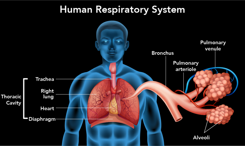
Breathing starts with the movement of air through yourmouth andnostrils. The human respiratory system begins at thetrachea. This is a strong tube reinforced by rings of cartilage. It starts at the back of your oral fissure and nose and and so splits into ii tubes calledbronchi.
Image - Text Version
Shown is an analogy of the human respiratory system and upper office of a male person human trunk. The parts of the respiratory system are opaque while the remainder of the torso is translucent.
Coming down from the oral cavity in the cervix area is a pink tube shaped object labelled equally the trachea. Branching off the bottom of the trachea are the dark pink and heavily veined lungs, one on each side of the body. The centre is likewise shown as a yellowish shape behind the lungs. At the base of operations of the lungs is a curved lite pink shape that is labelled as the diaphragm. The trachea, lungs, heart and diaphragm are labelled together as belonging in the thoracic cavity.
Coming off the left lung are some of the interior structures of the lung. These structures accept been significantly enlarged to brand them easier to place.
Directly coming off the lung is a pinkish, spaghetti-like construction that is labelled as the bronchus. The bronchus splits into branches, some of which stop in structures that resemble bunches of pink grapes. The grape-like objects are labelled as the alveoli. It is inside these structures that gas commutation takes place.
Wrapping effectually the bronchi and alveoli are 2 other thin, branching structures. The ones coloured red are labelled as pulmonary arterioles and the ones coloured blue are labelled as pulmonary venules. These are blood vessels that transport molecules to and from the lungs.
You tin can feel the cartilage rings in your trachea past running your fingers down the front of your neck. Tin can you feel the ridges? Those ridges are the cartilage rings! Each splits off many times to class smaller tubes called bronchioles. These bronchioles class a complex network of millions of little tubes that atomic number 82 to sacs called alveoli. This complex network of tubes and sacs forms the lungs.
Did you lot know?
Your left lung is smaller than your right lung. This is considering the left lung has to leave space for your eye!
Your lungs, too equally your heart, are located in your breast, also known as yourthorax. These organs are protected by a frame of basic called therib cage. Thediaphragm separates the thoracic crenel from the intestinal cavity, where the stomach and intestines are found. It is a big, dome-shaped muscle located beneath the rib cage.
Ventilation
Ventilation is the process by which air is pulled in and pushed out of the lungs. This is done by controlling the volume of thethoracic crenel, which is the space where the lungs are found. The main muscle responsible for ventilation is the diaphragm. Information technology is helped by theintercostal muscles, which are muscles between our ribs.
When you breathe in, the diaphragm and intercostal muscles contract. This increases the volume of the thoracic cavity and causes air to rush into the lungs. This phase of ventilation is calledinhalation.
Exhalation is when you breathe air out. During this stage, the diaphragm and intercostal muscles relax. This causes the space in the thoracic cavity to get smaller then air is forced out of the lungs.
Try taking a deep breath and imagining yous are filling your lungs completely full of air. If yous put your hands on your stomach, you can feel how far it moves out each time you breathe in.
The Gas Commutation Pathway
Permit's zoom in at a microscopic view and see how oxygen and carbon dioxide are transported and used in our bodies.
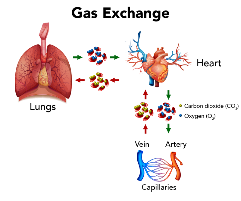
When an oxygen molecule is inhaled, it moves into the respiratory system through the mouth or nose. It so passes through the trachea, bronchi and bronchioles and into an alveolus. In the alveolus, the oxygen diffuses into theblood in the surrounding . Now the oxygen is in thecirculatory system.
are specialized cells that carry the gases in the claret. They practise this using a specialized protein calledhemoglobin. Within red blood cells, hemoglobin binds with molecules of oxygen and carbon dioxide. Hemoglobin relies on an iron molecule to make this happen. That'due south why it'south important to always accept enough fe in your nutrition. A deficiency in iron is calledanemia. This can cause fatigue equally well equally other more serious health issues.
You probably know that when iron rusts it looks reddish-orange. The same happens when hemoglobin binds to oxygen molecules. This is the reason why our claret looks red.
Did you know?
Some animals employ other molecules with their gas commutation proteins. Horseshoe venereal have a protein that binds with copper calledhemocyanin. This makes their blood expect blue. And information technology doesn't terminate there! Some animals accept imperial or even transparent blood!
Misconception Alarm
You might see bluish veins through your skin. But the claret in our veins is not blue. This is an effect of blueish light being the only 1 that can travel through our skin to our eyes!
Image - Text Version
Shown is a color illustration of the organs and cells involved in gas exchange. On the upper left is an illustration of the lungs. A light-green arrow on the right of the lungs points to red oval-shaped objects overlaid with small blue spheres. The ovals are cherry blood cells and the spheres represent oxygen molecules. From in that location another light-green arrow points to the heart. Below the heart, another dark-green pointer points to more blood cells with oxygen. A second light-green arrow points to a blood-red tube-shaped structure identified as an avenue. To the left of the artery is a similar blue structure identified as a vein. Between the avenue and vein are thin branching ruby-red and blueish lines identified as capillaries. To a higher place the vein is a blood-red arrow that points to blood cells that have yellowish spheres instead of bluish spheres. These spheres represent carbon dioxide molecules. To a higher place these claret cells is a red pointer pointing to the eye. To the left of the heart is a red pointer pointing to more claret cells with carbon dioxide. To the left of the blood cells is a red arrow pointing to the lungs.
The oxygenated blood is and so sent from the lungs to theeye which pumps it to the rest of the body through thearteries. Arteries branch out into all parts of the body and become smaller, until they becomecapillaries. Here oxygen diffuses from the blood to the trunk cells that demand it.
Diffusion is the motion of molecules from an area of high concentration to an area of depression concentration. Loftier concentration areas are where there are many of a certain type of molecule. Low concentration areas are the opposite.
Diffusion of oxygen from the claret to the body cells happens because the concentration of oxygen is higher in the blood than in the cells. Since molecules move from high to low concentration, oxygen moves from blood to cells.
Inside any trunk cell, oxygen is taken up bymitochondria. Some people call this thepowerhouse of the prison cell. In that location's a good reason for this. In the mitochondria, oxygen and sugars are used and produce energy for bodies. A waste product of thismetabolism is carbon dioxide.
Now, allow'southward follow a carbon dioxide molecule as it leaves the body. The concentration of carbon dioxide in the blood increases every bit cells produce it as waste. This causes it to diffuse into the blood. The carbon dioxide is carried past the red blood cells through theveins to the lungs. The lungs have a depression concentration of carbon dioxide compared to claret. This is why carbon dioxide diffuses into thealveoli of the lungs. From there it passes through bronchioles, bronchi, and the trachea to leave the trunk through the nose or mouth. This is the process ofexhalation.
The process of arresting oxygen and releasing carbon dioxide nosotros callgas exchange. All animals use a gas exchange to breathe. There are some differences, though, in the organs and molecules that make it happen.
Did yous know?
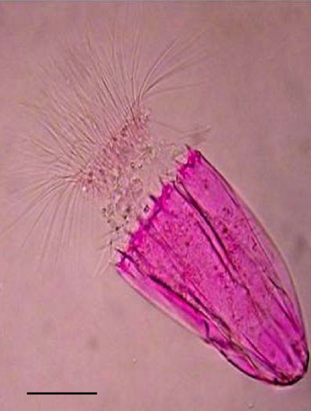
There is a group of tiny bottom-habitation marine organisms that live in an environment with no oxygen. It is believed that the Loricifera practise not accept mitochondria, and instead take a dissimilar blazon of organelle that can metabolize sugar without oxygen.
Epitome -Text Version
A pink-stained microscope image of a tiny organism resembling a closed umbrella pinnacle connected to a shaggy head of hair.
Respiratory Systems of Non-Mammalian Animals
Birds
The lungs of birds are organized very differently than those of humans. Unlike in homo lungs, where the air moves both in and out, in bird lungs, the air only flows in one direction. To help make this possible, birds have 9air sacs that environs the lungs. The air sacs permit for a continuous stream of air to menses through the lungs. It is important to note that gas exchange does not happen inside the air sacs.
For a complete respiratory cycle, birds crave two inhalations and ii exhalations.
- On thefirst inhalation air flows from the trachea to the rear air sacs.
- On theoutset exhalation air flows from rear air sacs to lungs.
- On thesecond inhalation air flows from lungs to front air sacs.
- On thesecond exhalationair flows from the front air sacs out through the trachea.
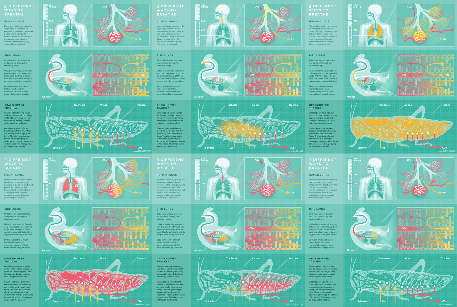
Image -Text Version
Human Lungs
Mammals inhale by moving the diaphragm to lower the air pressure in the chest crenel and pull air into the lungs. The human chest cavity is always at a lower pressure level than the exterior environment (usually 760mmHg at body of water level).
Bird Lungs
Birds accept air sacs that store and pump air through the stationary lungs.
Different in mammals, air flows in only one direction through bird lungs. With the help of the air sacs, this allows birds to have in oxygen even during exhalation.
Birds can exhale at much higher elevations than mammals because of their more than efficient lung structure.
Grasshopper Trachea
Grasshoppers have no lung and exercise not apply their circulatory organisation to move oxygen. They send air directly to tissue cells using tracheal tubes.
Grasshoppers apply dissimilar animate methods when they are resting, alarm, hopping, or flying. The alert grasshopper shown hither is pumping its abdomen to change the volume of its air sacs. This helps pump air through the trachea.
The bird respiratory organization is far more efficient than that of mammals. The continuous flow of oxygen is important to birds equally they need a lot of energy to fly. Some other advantage of the air sacs is that they make birds less heavy!
Reptiles
The respiratory system of reptiles is like to that of humans. I major exception is that almost reptiles, except for members of the crocodile family, do not have a diaphragm. They have evolved different means to inflate their lungs.
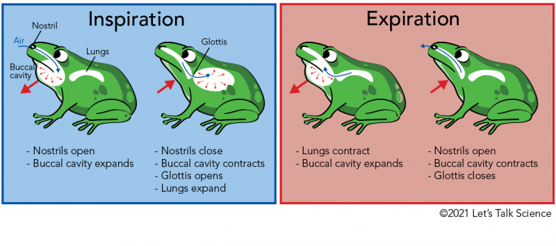
Many reptiles and amphibians use their throat muscles to "gulp" air in a process chosenbuccal pumping.
Image - Text Version
Shown is a colour illustration showing the four-step process of inspiration and expiration in a frog.
On the left side is a blue rectangle with the title, "inspiration." In the rectangle are 2 drawings of frogs. The frog on the left has several trunk parts labelled. These include the nostrils, which are located above the rima oris, the lungs, which are located in the main part of the body, and the buccal cavity which is located in the throat area. In this outset stage of inspiration, a blueish arrow shows how air moves from outside the frog, through the nostril and into the buccal crenel. Red arrows show that when this happens, the buccal cavity expands outwards. The frog on the right shows what happens in the next stage of inspiration. The nostril is shown as closed, a blueish arrow points to air moving past a part identified as the glottis and into the lungs. One red arrow points to the buccal cavity which has contracted. Another cerise arrow shows that the lungs are expanding.
On the correct side is a pink rectangle with the title, "expiration." Again, there are two drawings of frogs. The frog on the left illustrates what happens during the first stage of expiration. A blue arrow shows how air moves from the lungs and through the glottis. Red arrows bear witness that when this happens, the buccal cavity expands outwards. The frog on the correct shows what happens in the second stage of expiration. The nostril is shown as open, a blue arrow points to air moving out through the nostril. One ruddy arrow points to the buccal cavity which has contracted. At this phase the lungs have contracted.
There are four steps in buccal pumping.
- Commencement, air is let in through thenostrils. Thebuccal cavity, which is connected to the mouth, expands.
- When the buccal cavity is full, the nostrils shut and airways to the lungs open. The opening to the airway is called theglottis. Then, the fauna uses muscles to push air into the lungs.
- When exhaling, first the lungs contract which pushes air into the buccal cavity. The buccal cavity once again expands.
- Finally, the glottis closes, the buccal crenel contracts and the nostrils open up.
Turtles use many unlike methods to breathe. Some move their legs into and out of their shells to aid inflate and deflate their lungs. Others have large muscles that wrap effectually the lungs to command the book of the thoracic cavity. Finally, some aquatic species can fifty-fifty utilise theircloacato breathe. The cloaca is the external opening used for solid, liquid excretions and access to the reproductive arrangement. In mammals, there are different openings for these things. Cloacal breathing is by and large used when turtles hibernate underwater. It is considered to exist a type of respiration, which is gas substitution that happens through the skin.
Did you know?
Ocean turtles can hold their breath for up to 10 hours!
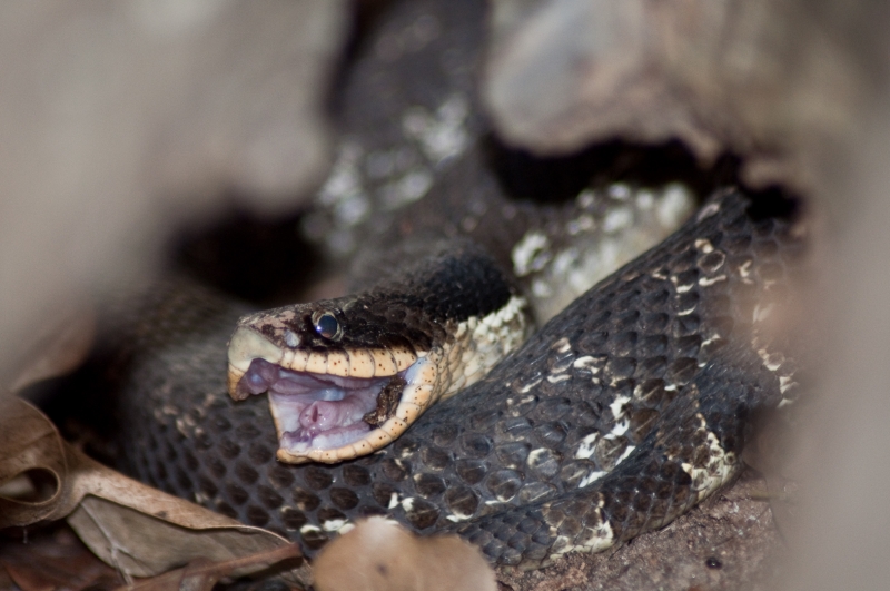
Another interesting item of most reptiles is that they lack a separation betwixt their nose and mouth. This forces them to stop animate to exist able to swallow. To deal with the long time it takes to swallow their prey, snakes have a long extension of the trachea.
Paradigm - Text Version
Shown is a colour photo of a snake. The snake is night brown with small beige patches here and there. The area around the mouth is a pale orange. Merely the head and a few coils of the torso are visible through some blurry leaves. The serpent's oral fissure is wide open. Within the pale pink mouth is a pink tube-shaped structure. This is the snake's trachea.
Amphibians
Amphibiansare animals that have adapted to living both on land and in water. This group includes frogs, toads, salamanders and newts. Amphibians are interesting because they start off equally aquatic larvae before they move on state as adults. As they abound and move from living in water to living on land, their respiratory systems change.
In their juvenile forms, amphibians acceptgills that let them breathe underwater, just like fish. As they abound to their adult grade, the gills disappear and lungs develop to replace them. Some species of salamanders remain aquatic, even so, and go along their gills all through their lives. We telephone call keeping characteristics of the larval stageneoteny.
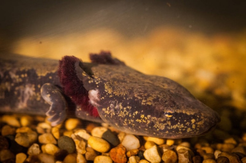
Neoteny is easily seen in axolotl (pronounced axe-o-lot-sick), a popular aquarium species from Mexico. In Canada, our largest salamander species is the mudpuppy. Information technology is easy to recognize because it has reddish gills on each side of its head.
Image - Text Version
Shown is a colour photograph of a mudpuppy. Just the head, forepart right leg and a pocket-sized portion of the body is visible. The mudpuppy is mainly a purplish-grey colour. It also has golden splotches all over its body. It has a wide, apartment head with modest gold and blackness eyes. Just behind the head are projections, similar to ears, that accept dark red feathery edges. These are their gills.
In addition to buccal pumping, amphibians as well apply . This ways that the gas exchange happens through the beast's peel. Many other animals take cutaneous gas exchange, fifty-fifty humans, simply in small quantities! Amphibians rely on this procedure much more. The lungless salamander, as its proper noun implies, almost exclusively uses its peel to exhale. This animate method requires having thin skin that can be kept wet, which many amphibians have. Since their breathing happens through their peel, it is all-time not to handle amphibians. Not simply tin yous affect their animate, you tin can as well transfer germs from your skin to theirs.
Fish
The respiratory system in fish is very different from the respiratory systems ofanimals. Like frog larvae and mudpuppies, fish have gills. Gills are similar to lungs in that they have branches that split.Gill arches branch to gradegill filaments. On the gill filaments aregill lamellae, which is where gas exchange occurs. Hither oxygen dissolved in water diffuses into the blood and carbon dioxide diffuses into the water.
In , a bony plate called anoperculum covers and protects the fragile gills. Incartilaginous fishes, such as sharks and rays, the gills are located near their 5 to 7 gill slits.
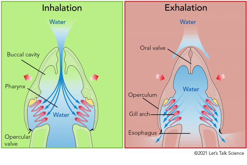
During the inhalation phase, fish pull water in through theiroral valve to their buccal cavity. From at that place the water is pushed towards their gills. Theopercular valve is closed at this indicate. During the exhalation phase, the oral valve is closed and the opercular valve is opened. Water passes over the gills and out through the opercular valve.
Image - Text Version
Shown is a color analogy of the processes involved in fish inhalation and exhalation.
On the left side is a stake green rectangle with the title, "Inhalation." Below it is a cutaway view of a fish's head. At the height end is the oral fissure. It is fairly pointed and has a narrow opening which connects with the larger buccal cavity. Pinkish arrows on either side of the head point that the fish is pushing its gills outwards. When this happens, blue arrows signal that water comes in through the oral valve and into the buccal cavity and and so on to the pharynx at the dorsum of the head. The h2o then heads towards the gill arches, which are light pink two-pronged structures. Beyond the gill arches is an opening with a modest flap in the airtight position. This is the opercular valve. Some water too passes into the fish's esophagus at the back of the caput.
On the right side is a pink rectangle with the title, "Exhalation." Below it the aforementioned cutaway view of a fish's head equally in the previous image. Pink arrows on either side of the head indicate that the fish is pulling its gills inwards. At this point the oral value at the mouth is airtight. Bluish arrows signal that some water bypasses the fish's caput. Other bluish arrows within the pharynx area indicate that the h2o is passing over the fill arches. Some of the water is also passing through the at present open up opercular valve and out to surrounding h2o. About water does non pass into the esophagus at this phase.
Some fish have specialized muscles that let them to pump the h2o over their gills. Fish that are very active use a type of ventilation calledram ventilation. Moving forrad forces water to movement over the gill arches so that oxygen and carbon dioxide can be exchanged. Sharks are a type of fish that use ram ventilation. This is why some species of sharks tin can actually drown if they stop moving!
Did you know?
Sharks and rays bring water into their buccal cavities through an actress opening behind the eye called aspiracle. Spiracles are likewise used by insects for respiration.
Source: https://letstalkscience.ca/educational-resources/backgrounders/respiratory-system-in-vertebrate-animals
Posted by: southwoodperaweltake.blogspot.com

0 Response to "How Do Animals Get Rid Of Carbon Dioxide"
Post a Comment